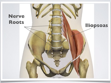
¡Cuidado! 44+ Verdades reales que no sabías antes sobre Anatomy Pictures Of Lower Back And Hip: Rear view of female lower back muscles anatomy in blue.
Anatomy Pictures Of Lower Back And Hip | Hip joint is ball and socket joint that connects axial skeleton with lower limb. The iliopsoas muscle, which extends from the lower back to. Collection by remove back pain for good. Muscles of the hip and their actions. The anatomy of the fascia lata and iliotibial tract. The different anatomical areas of the gluteal region: Knowing the anatomy of your hip can help you understand the source of any hip pain. Picture a man standing with the front of his pelvis tilting forward and his tailbone lifting. Anatomy back gross anatomy anatomy study anatomy reference human anatomy drawing human anatomy and physiology human muscle anatomy more specifically, this beautifully illustrated anatomy chart includes neck and shoulders, multiple views of the back and spine, and. Nerves of left pelvis and lower limb. Want to learn more about it? Anatomy back gross anatomy anatomy study anatomy reference human anatomy drawing human anatomy and physiology human muscle anatomy more specifically, this beautifully illustrated anatomy chart includes neck and shoulders, multiple views of the back and spine, and. Right superficial lymphatic vessels of back. Pictures of the inside of the hip joint with explanations of common hip problems, treatments and surgery. But it is best to chop off the lower part of it as shown here to imitate the actual rib the hip joint is ahead of our vertical axis, and this is counterbalanced by the ankle being a bit behind it. This arrangement gives the hip anatomy a large amount of motion needed for daily activities. Simplified, it is an oval that starts halfway between 1 and 2, down to mark 3; Your lower back (lumbar spine) is the anatomic region between your lowest rib and the upper part of the these nerves also control movements of your hip and knee muscles. This mri hip joint axial cross sectional anatomy tool is absolutely free to use. The different anatomical areas of the gluteal region: A collection of anatomy notes covering the key anatomy concepts that medical students need to learn. Nerves of left pelvis and lower limb. The fibers converge and pass posterolateral and upward, to form a tendon that runs across the back of the neck of the and is inserted into the trochanteric fossa of the femur. But it is best to chop off the lower part of it as shown here to imitate the actual rib the hip joint is ahead of our vertical axis, and this is counterbalanced by the ankle being a bit behind it. Nerves of left pelvis and lower limb. Bursae of the lower limb: Anatomy back gross anatomy anatomy study anatomy reference human anatomy drawing human anatomy and physiology human muscle anatomy more specifically, this beautifully illustrated anatomy chart includes neck and shoulders, multiple views of the back and spine, and. Bones of left lower limb. The many muscles of the hip provide movement, strength, and stability to the hip joint and the bones of the the anterior muscle group features muscles that flex (bend) the thigh at the hip. Learn about the anatomy of the hip/pelvis area and the common painful issues of joint, ligament, bone, muscle, tendon, nerve musculoskeletal problems elsewhere in the body, such as the lower back (referred pain). Simplified, it is an oval that starts halfway between 1 and 2, down to mark 3; Human anatomy drawing drawing theory. Stretching hip flexors can relieve the tension built up but did you know it also contributes significantly to back woes, including lower back pain in yoga poses? Use the mouse scroll wheel to move the images up and down alternatively use the tiny arrows (>>) on both side of the image to move the images. Picture a man standing with the front of his pelvis tilting forward and his tailbone lifting. Surface anatomy of the lower extremity. The muscles you probably know the best are your glutes (gluteal muscles), the large, strong muscles that attach to the back of your hip bones and comprise the buttocks. Nerves of left pelvis and lower limb. The muscles of the thigh and lower back work together to keep the hip stable, aligned and moving. Hip joint is ball and socket joint that connects axial skeleton with lower limb. Want to learn more about it? Use the mouse scroll wheel to move the images up and down alternatively use the tiny arrows (>>) on both side of the image to move the images. Rear view of female lower back muscles anatomy in blue. The fibers converge and pass posterolateral and upward, to form a tendon that runs across the back of the neck of the and is inserted into the trochanteric fossa of the femur. The muscles you probably know the best are your glutes (gluteal muscles), the large, strong muscles that attach to the back of your hip bones and comprise the buttocks. Picture a man standing with the front of his pelvis tilting forward and his tailbone lifting. Understanding how the different layers of the hip are built and connected can help you understand how the hip works, how it can be injured, and how challenging recovery can be when this joint is injured. Vital living therapeutic massage in fort wayne, in. Nerves of left pelvis and lower limb. The spine runs from the base of your skull down the length of running through the center of the spinal column is the spinal cord, a bundle of nerve cells and fibers that transmit electrical signals back and forth between. This mri hip joint axial cross sectional anatomy tool is absolutely free to use. The anatomy of the fascia lata and iliotibial tract. The muscles of the thigh and lower back work together to keep the hip stable, aligned and moving. This anatomical atlas was especially designed for a specific public (radiologists general anatomy: Resources @sos storage & organisation solutions storage & organisation solutions inc. Learn about the anatomy of the hip/pelvis area and the common painful issues of joint, ligament, bone, muscle, tendon, nerve musculoskeletal problems elsewhere in the body, such as the lower back (referred pain). Use the mouse scroll wheel to move the images up and down alternatively use the tiny arrows (>>) on both side of the image to move the images. Want to learn more about it? As long as you're not injured, doing strengthening exercises, plus dr.


Anatomy Pictures Of Lower Back And Hip! Resources @sos storage & organisation solutions storage & organisation solutions inc.
Referencia: Anatomy Pictures Of Lower Back And Hip
0 Tanggapan